Sixty Years of Lung Cancer Screening: What Actually Worked
By ELESSAR
Sixty Years of Lung Cancer Screening: What Actually Worked
Lung cancer kills more people than any other cancer, largely because it is usually found too late. A new review follows six decades of attempts to catch it early, and explains why one method finally succeeded where others failed.
Lung cancer causes more deaths worldwide than any other malignancy, and the reason is painfully simple. By the time it causes symptoms, a cough that will not settle, breathlessness, weight loss, it has often already spread. A tumour caught while it is still small and confined to one part of the lung can frequently be removed with surgery. One that has reached the lymph nodes or distant organs rarely can. So for sixty years, researchers have chased the same idea: find the cancer before the person feels ill, when treatment still has a chance of working. A recent review published in Academic Radiology traces that long effort, and it reads less like a triumph than a lesson in how hard early detection actually is.
The first serious attempts, beginning in the 1960s, relied on two tools. One was the plain chest X-ray, which can show a shadow in the lung. The other was sputum cytology, in which a sample of coughed-up mucus is examined under a microscope for abnormal cells. Both seemed reasonable. Both were tested in large studies. And both delivered a result that took years to accept: they found more cancers, and even shifted some diagnoses to earlier stages, yet they did not reduce the number of people dying from lung cancer. The review explains why this can happen. A test that detects slow-growing tumours that would never have caused harm inflates the count of early cancers without saving lives, a distortion known as overdiagnosis. And a test can appear to lengthen survival simply by moving the diagnosis date earlier, without changing the day a person actually dies. Catching a cancer sooner is not the same as changing its outcome.
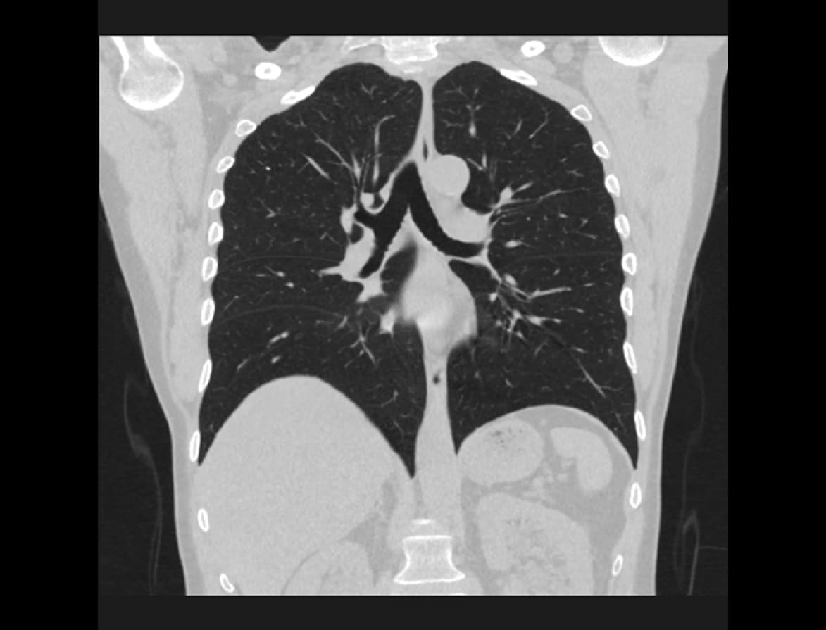
The turning point came with computed tomography, or CT, which builds a detailed three-dimensional image of the lungs and can reveal nodules far smaller than an X-ray shows. The concern was radiation, so researchers developed low-dose CT, which uses a fraction of the radiation of a standard scan. The review draws on the two large randomised trials that tested it, meaning studies where people were assigned by chance to be screened or not, the strongest design for judging whether an intervention truly helps. In the American trial, heavy smokers and former smokers who received annual low-dose CT scans had a meaningfully lower rate of death from lung cancer than those given chest X-rays. A large European trial later reported a similar reduction compared with no screening. For the first time, a screening method had passed the test that X-rays and sputum failed: fewer people died of the disease. This is why low-dose CT is now recommended in many countries for people at high risk because of a long, heavy smoking history.
The review is careful not to present screening as free of harm. The most common problem is the false alarm. Low-dose CT is sensitive enough to spot many small nodules, and the large majority of them are harmless scars or benign growths. Each one, though, can trigger anxiety, repeat scans and sometimes an invasive procedure such as a biopsy to rule out cancer, and those procedures carry their own small risks. Overdiagnosis remains a genuine concern even with CT: some detected cancers may never have threatened the person's life, yet once found they are usually treated. There is also the cumulative radiation of yearly scans, and the reality that screening only helps if it reaches the right people and is followed up properly. The benefit is real, but it is a balance, and it is strongest in those whose smoking history puts them at genuinely high risk.
Looking forward, the review describes a shift toward what it calls a precision era. Rather than defining eligibility mainly by age and pack-years of smoking, researchers are developing risk models that combine many factors to identify who would benefit most. Blood-based biomarkers, molecular signals that might flag cancer earlier or tell a dangerous nodule from a harmless one, are under active study, though they are not yet ready to replace imaging. Artificial intelligence is being trained to read scans and help distinguish nodules that need attention from those that do not. These directions are promising rather than proven; the review presents them as the current frontier, not as established practice.
Sources
- Sixty Years of Lung Cancer Screening: From Sputum Cytology to Low-Dose CT and the Emerging Precision Era.Sixty Years of Lung Cancer Screening authorship (Academic Radiology) · 2026 · Academic RadiologyDOI 10.1016/j.acra.2026.08.052PMID 42674935